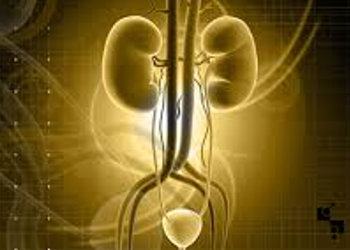
 Calculus (stone) formation in the urinary tract is the third in order of frequency disease in humans after urinary tract infections and prostate conditions. Such stones in the urinary tract appear at any age and sex, in every race and country and are known since ancient times.
Men are affected more often than women at a ratio of 2.5:1, with a higher incidence at the age of 30 for men, while for women the appearance of such stones is more common between the ages of 35 and 55 years. Moreover, it has been found that the probability of forming a new stone one year after the initial episode reaches 10%, and within 5-7 years the recurrence rate is 50%.
Today it is considered that the occurrence of calculi is due to hereditary and genetic defects. However, studies have shown that in 25% of patients who reported calculus formation there is also a presence of calculi in other family members, which is probably due to the same eating habits and living conditions.
The incidence of calculi shows geographical distribution meaning that different geographical zones are associated with different calculi rates. This is due to the difference in climatic conditions and the content of trace elements in drinking water e.g. calcium and magnesium.
Increased frequency of calculi is also present in people who, because of their way of working, have limited mobility, while improving the living conditions, namely increasing consumption of animal protein and animal fat, increased the incidence of urolithiasis. It has to be noted that the prevalence of lithiasis increased steadily during the 20th century, except for the periods of the 1st and 2nd World War, which was attributed to the change of diet associated with reduced intake of animal protein during the wars.
Finally, taking plenty of fluids is another important dietary factor in preventing calculi. Small intake or large fluid losses create favorable conditions for the occurrence of urolithiasis.
What kind of symptoms are experienced in patients with urolithiasis?
Calculus formation in the urinary tract appears clinically as acute pain in the lumbar region and the lower quadrant of the abdomen. It is characteristic of the patient to keep moving continuously unable to find relief in any position. As the stone approaches the bladder, the pain characteristics change and expand to the area of the bladder, while there is a strong urinary urgency as well as frequency. As the tension of the stomach and the kidney remains the same, the pain is most often accompanied by nausea and vomiting.
How is the diagnosis of lithiasis performed?
The first step to be taken in order to diagnose lithiasis is to ask for a simple radiography of the kidneys, ureters and bladder. The ultrasound is a rapid, easy and safe test that helps to diagnose the presence/absence of calculi because it provides additional information. In some cases when there is a diagnostic problem, intravenous urography (pyelography) is recommended.
What is the treatment of calculi?
Treatment of calculi is twofold, initially it focuses on the alleviation of pain and then on the removal of the stone. The best and final treatment is either the automatic expulsion of the stone or its surgical removal.
The decision on the type of treatment of a stone depends mainly on its size and secondly, on its localization. In about 85% of the cases, the diameter of the stones is less than 5 mm and they are usually expelled automatically. The chances of the stones being expelled automatically increase the lower they are located, i.e. as close to the bladder. In the absence of spontaneous expulsion of the stone, the time limit of four weeks is considered a reasonable waiting time, since the kidney is not in danger during that time period, as it has been found that detectable damage in a previously normal kidney occurs after 4 weeks of complete obstruction.
When is medical intervention required for the removal of the stones and how is it done?
Treating calculi consists in removing the stone. For this purpose, in our modern era of minimally invasive treatments, surgical removal of stones is rarely performed, since any urologist may utilize a wide array of effective and also less invasive methods. Such methods are extracorporeal lithotripsy, ureteroscopy and percutaneous nefrolithotripsy. It is noted that extracorporeal lithotripsy and percutaneous nefrolithotripsy have contraindication in pregnancy.
Extracorporeal lithotripsy
Extracorporeal shockwave lithotripsy appeared in 1980 and brought a true revolution in the treatment of calculi. It works best with stones of the kidney and with the initial part of the ureter. Almost all kinds of stones are treated by this method.
Ureteroscopy and intracorporeal (endoscopic) lithotripsy
Ureteroscopy is a method in which a long, thin body, the ureteroscope, is inserted, while being visually guided, through the urethra and bladder into the ureter to the point where the stone is located. Through the ureteroscope, the lithotripters are introduced whose function is to crush the stone, while special pliers which are also introduced through the ureteroscope, grasp and remove the remaining stone pieces.
Percutaneous nefrolithotripsy
Percutaneous lithotripsy is a method with extremely high success rates. It is primarily indicated for those stones that can not be treated by extracorporeal lithotripsy. Such cases are stones larger than 2.5 cm., coral stones, hard stones that are highly resistant to extracorporeal lithotripsy and stones in patients with deformity of the skeleton or major obesity.
In conclusion, urolithiasis is a common disease of the urinary tract. The possibility of using high-tech applications has introduced a new dimension in the treatment of calculi. Injuring the patient as little as possible is always a priority. Thus seeking a passage to the urinary system through its natural pathway, such as the urethra and ureters or the pathway that is formed by an organ's medical puncturing such as the kidney, is always preferred. The creation of an appropriate entry to the urinary tract, opens up the possibility of performing a series of operations in the kidney, ureter and bladder.